CDKN2A/p16INK4a Rabbit mAb
目录号: F1180
仅在部分组织和细胞系中表达,如宫颈癌、乳腺癌等肿瘤组织;在正常组织中几乎不表达。如需获取不同样本的表达情况请点击“样品处理数据示例表”。
抗体应用:
反应性:
-
 Lane 1: Hela, Lane 2: HEK-293T, Lane 3: Saos-2
 -
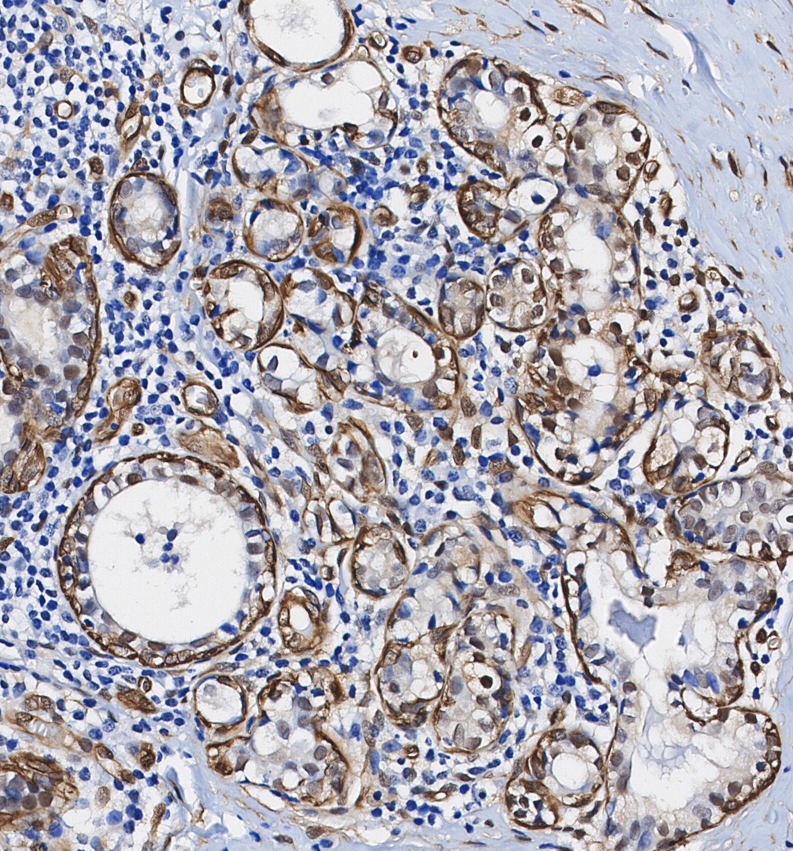 Immunohistochemical analysis of formalin fixed paraffin embedded human Breast Cancer tissue with F1180 at 1/100 dilution.
操作要点
仅在部分组织和细胞系中表达,如宫颈癌、乳腺癌等肿瘤组织;在正常组织中几乎不表达。
WB
湿转参考条件:200 mA, 60 min,建议使用 0.22 μm PVDF膜。
CDKN2A/p16INK4a蛋白为小分子蛋白(17kda),推荐使用 0.22 μm PVDF膜,湿转条件200 mA, 60 min。
WB实验中建议添加阳性对照组(HeLa、HEK-293等),用于验证实验体系可靠性。CDKN2A/p16INK4a蛋白存在多个异构体,在WB实验中可能检测到多条带现象。
使用信息
| 稀释比例 |
|---|
|
| 抗体应用 |
|---|
| WB, IP, IHC, FCM |
| 反应性 |
|---|
| Human |
| 抗体类型 |
|---|
| Rabbit |
| 储存液配方 |
|---|
| PBS, pH 7.2+50% Glycerol+0.05% BSA+0.01% NaN₃ |
| 储存条件(自收到货起) |
|---|
| –20°C (avoid freeze-thaw cycles), 2 years |
|
预测分子量
|
|---|
|
17 kDa
|
| 阳性对照 | Human testis; Human normal breast; Human cervical carcinoma; Human CDKN2A full-length recombinant protein; HeLa; HEK-293; Saos-2 |
|---|---|
| 阴性对照 | MCF7 |
样品处理数据示例
| 样品 | 处理情况 |
| A549 | Low Expression |
| HESCs | Low Expression |
| A498 | Low Expression |
| 点击查看更多样品数据 | |
*该蛋白在不同的人源细胞、组织内的表达量预测请参考:http://www.proteinatlas.org
实验方法
| WB |
|---|
实验步骤: 样品制备
1. 组织样品:破碎组织,加入适量冰冷的 RIPA/NP-40 Lysis Buffer (含蛋白酶抑制剂Cocktail),低温匀浆或置于冰上超声裂解样品,静置30 min。 2. 贴壁细胞样品:吸去培养基, 将细胞转移至EP管中。用冰冷的 PBS 清洗细胞 2 遍。加入适量冰冷的 RIPA/NP-40 Lysis Buffer (含蛋白酶抑制剂Cocktail),超声破碎,冰上静置裂解 30 min。冰上静置裂解 5 min。 3. 悬浮细胞样品:将培养基转移至离心管中离心,弃上清,用冰冷的 PBS 清洗细胞 2 遍。加入适量冰冷的 RIPA/NP-40 Lysis Buffer (含蛋白酶抑制剂Cocktail),冰上静置裂解 5 min。 4. 将所得匀浆液/裂解液置于离心机中 4°C 离心 15 min,收集上清液;
5. 取少量裂解液测定蛋白质浓度;
6. 加入蛋白上样缓冲液,将 20 µL样品在 95~100°C加热 5 min,冰上静置冷却后离心 5 min。
电泳分离
1. 根据所提蛋白的浓度,将适量蛋白样品和 Marker 上样至 SDS-PAGE 凝胶。建议分离胶(即下层胶)浓度:10 %。 SDS-PAGE 分离胶浓度选择参照表 2. 电源调 80 V, 30 min。然后电源调 110 V~150 V,观察 Marker,待蛋白所在的预染蛋白 Marker 指示带得到合适的分离后,即可停止电泳。(注意电泳时电流切勿过大,如超过 150 mA 会导致温度上升,容易影响跑胶结果。如无法避免采用大电流,可对电泳槽使用冰浴降温。)
转膜
1.拿出电转槽,把夹子和耗材浸泡在预冷电转液中;
2.用甲醇活化 PVDF 膜 1 min,用转移缓冲液冲洗;
3.按照“夹子黑边-海绵-滤纸-滤纸-胶-PVDF膜-滤纸-滤纸-海绵-夹子白边”的顺序安装好; 4.将蛋白电转移至 PVDF膜上。(推荐采用 0.22 µm PVDF 膜) PVDF 膜孔径规格选择参照表 湿转法参考条件:200 mA, 60 min。
(注意电转条件可根据蛋白大小适当调节,分子量大的蛋白适宜采用大电流,并延长转膜时间,但需要确保电转槽始终处于低温的环境中,避免凝胶融化。)
封闭
1. 电转移后,室温下用 TBST 洗膜 5 min;
2. 在封闭液中将膜孵育 1 h,室温;
3. 用 TBST 洗膜 3 次,每次 5 min。
抗体孵育 1. 用 一抗稀释液配制一抗工作液(建议一抗稀释比 1:2000),4°C 条件下与膜轻柔摇晃孵育过夜; 2. 用 TBST 洗膜 3 次,每次 5 min;
3. 在封闭液中加入二抗,室温条件下与膜轻柔摇晃孵育 1 h;
4. 孵育结束后,用 TBST 洗膜 3 次,每次 5 min。
显色
1. 加入配制好的 ECL 发光底物(或根据二抗选择其他显色基质)混合均匀;
2. 与膜孵育1 min,去除多余底物(需保持膜湿润),置于显影仪中进行曝光。
|
| IF |
|---|
| IHC |
|---|
实验步骤:
脱蜡/补液
1. 脱蜡/水合切片:
2. 将切片在二甲苯中孵育 3 次,每次 5 分钟。
3. 将切片在 100% 乙醇中孵育两次,每次 10 分钟。
4. 将切片在 95% 乙醇洗涤液中孵育两次,每次 10 分钟。
5. 用 dH2O 清洗切片两次,每次 5 分钟。
6.抗原修复:对于柠檬酸盐:将载玻片浸入 1X 柠檬酸盐暴露溶液中,在微波炉中加热,直至开始沸腾; 在亚沸温度 (95°-98°C) 下继续煮 10 分钟。 在工作台上冷却玻片 30 分钟。
染色
1. 用 dH2O 清洗切片 3 次,每次 5 分钟。
2. 将切片在 3% 过氧化氢中孵育 10 分钟。
3. 用 dH2O 清洗切片两次,每次 5 分钟。
4. 在洗涤缓冲液中洗涤切片 5 分钟。
5. 用 100–400 µl 封闭液在室温下封闭每个切片 1 小时。
6. 除去封闭液,并向每个切片中添加 100–400 µl 一抗稀释液。 4°C 孵育过夜。
7. 去除抗体溶液,用洗涤缓冲液洗涤切片 3 次,每次 5 分钟。
8. 用 1-3 滴所需的 HRPA 覆盖切片。 在加湿室中室温孵育 30 分钟。
9. 用洗涤缓冲液洗涤切片 3 次,每次 5 分钟。
10. 使用前将 DAB 显色剂浓缩液加入 DAB 稀释液中并充分混合。
11. 在每个切片上涂抹 100–400 µl DAB 并密切监测。 1-10 分钟通常可提供可接受的染色强度。
12. 将载玻片浸入 dH2O 中。
13. 如果需要,用苏木精复染切片。
14. 用 dH2O 清洗切片两次,每次 5 分钟。
15. 切片脱水:95%乙醇孵育切片两次,每次 10 秒; 在 100% 乙醇中重复,孵育切片两次,每次 10 秒; 在二甲苯中重复,孵育切片两次,每次 10 秒。
16. 用盖玻片和封固剂封固切片。
|
生物描述
| 特异性 |
|---|
CDKN2A/p16INK4a蛋白的表达水平可能因样品类型而异。该蛋白通常在一些肿瘤组织中表达,如宫颈癌、乳腺癌组织等,在正常组织中几乎不表达。此外,也仅在某些特定细胞系中才有表达。 |
| 蛋白定位 |
|---|
| 细胞质,细胞核 |
| Uniprot ID |
|---|
| P42771 |
| 克隆号 |
|---|
| P15P14 |
| 别名 |
|---|
| CDKN2A/p16,p16 INK4A |
| 背景 |
|---|
P16INK4a (p16) 是一种由 CDKN2A 基因编码的细胞周期蛋白依赖性激酶抑制剂,它通过抑制 CDK4/6 来阻止 E2F 转录因子的激活。CDKN2A 有三个外显子:外显子 1(150 个碱基对,编码氨基酸 1-50)、外显子 2(307 个碱基对,编码氨基酸 51-153)和外显子 3(11 个碱基对,编码氨基酸 153-156)。CDKN2A 基因可以通过 5'-CpG 岛甲基化在各种癌症中失活,包括肺癌、神经胶质瘤和头颈癌。CDKN2A 表达的失调可以通过缺失、剪接、错义和无义突变以及甲基化发生。 p16 蛋白通过与 D-细胞周期蛋白竞争结合其催化亚基来抑制 CDK4 和 CDK6,从而通过视网膜母细胞瘤蛋白 pRb 将 p16INK4a 与细胞周期调控联系起来。 |
| 参考文献 |
|---|
技术支持
在订购、运输、储存和使用我们的产品的任何阶段,您遇到的任何问题,均可以通过拨打我们的热线电话400-668-6834,或者技术支持邮箱tech@selleck.cn,直接联系到我们。我们会在24小时内尽快联系您。
* 必填项
